Potential Severity
Subdural abscess spreads rapidly. Emergency surgical drainage is life-saving.
Intracranial epidural and subdural abscesses are rare. They usually result from spread of infection from a nidus of osteomyelitis after neurosurgery from an infected sinus (in particular the frontal sinus), or less commonly, from an infected middle ear or mastoid. In infants, subdural effusions may complicate bacterial meningitis; however, unlike the form seen in adults, they rarely require drainage. The bacteria causing these closed-space infections reflect the primary site of infection. S. aureus is most common, followed by aerobic streptococci. Other pathogens include S. pneumoniae, H. influenzae, and gram-negative organisms. Anaerobes such as anaerobic streptococci and B. fragilis can also be associated with this infection. Patients with sinusitis and chronic mas-toiditis often have polymicrobial abscesses.
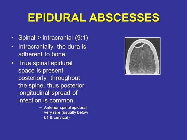
Epidural abscesses form between the skull and the dura. Because the dura is normally tightly adherent to the skull, this infection usually remains localized and spreads slowly, mimicking brain abscess in its clinical presentation. On exam, localized erythema, swelling, and tenderness of the subgaleal region may be seen. Subdural empyema in the cranial region progresses much faster than epidural abscess does, usually spreading rapidly throughout the cranium.
About Epidural and Subdural Intracranial Abscess

- Associated with frontal sinusitis, mastoiditis, and neurosurgery.
- Staphylococcus aureus are a common cause; otherwise, microbiology is similar to that in brain abscess.
- Epidural abscess progresses slowly, requires surgical drainage.
- Subdural abscess spreads quickly.
- Often mimics meningitis.
- Lumbar puncture is contraindicated; use computed tomography scan or magnetic resonance imaging emergently.
- Requires immediate drainage.
- Mortality ranges from 14% to 18%.
Patients appear acutely ill and septic. They complain of severe headache that is localized to the site of infection, and nuchal rigidity commonly develops, suggesting the diagnosis of meningitis. Within 24 to 48 hours focal neurologic deficits are noted, and half of these patients develop seizures. Lumbar puncture is contraindicated because of the high risk of brain stem herniation. A computed tomography scan with contrast should be performed, and in most instances, the images demonstrate the abscess and the overlying osteomyelitis, sinus infection, or mastoiditis. In early epidural or subdural abscess, magnetic resonance imaging scan is capable of detecting early cortical edema and smaller collections of inflammatory fluid. In patients suspected of having early disease, whose computed tomography scan is negative, an magnetic resonance imaging scan should be performed.
Subdural empyema is a neurosurgical emergency. Immediate drainage is required to prevent death from cerebral herniation. Exploratory burr holes and blind drainage have been life-saving in rapidly progressing cases. Antibiotic therapy should be instituted immediately. The same regimens recommended for brain abscess are used. The mortality from subdural empyema remains high at 14% to 18%, the prognosis being especially poor in patients who are comatose. Epidural abscess is less dangerous, but also requires surgical drainage. Mortality is low; however, if left untreated, this infection can spread to the subdural space.


